As there are a wide range of Coral types, I am just going to focus on the general anatomy and focus in on the anatomy of the Rugosa, Tabulata and Scleractinia orders.
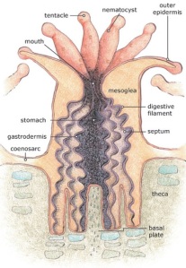
Corals belong to the Phylum Cnidaria which also includes jellyfish and sea anemones. Corals are diploblastic, meaning they have two layers of cells; the endoderm (inner layer) and the ectoderm (outer layer). A key anatomical feature of cnidarians is the presence of nematocysts. These are stinging capsules which help in defence and predation. The coral body structure is radially symmetrical and has one main form; the polyp stage which is sessile. The polyp anatomy is cylindrical and attached to the substrate with tentacles at the oral end (Campbell & Reece, 2005). (www1) described the polyp as multicellular with specialised cells. Corals have limited organ development and do not possess a central nervous system. However corals do have a gastrovascular cavity and a ring of tentacles.
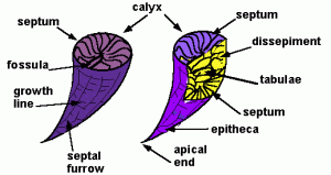
Coral skeletons are made up of calcium carbonate (CaCO3) present in one of two mineral forms, either calcite or aragonite. Corals whose skeleton are made of calcite are much more likely to be preserved in the fossil record than those with aragonite skeletons as aragonite is a much less stable mineral. The general coral skeleton contains the theca (wall), the septa (radiating plates), tabulae and dissepiments which are horizontal strengthening structures (www2) (see fig. 1). (www1) stated that hard corals have a calyx (cup) which the coral polyp is located in. The bottom of the calyx is known as the basal plate. The stomach has a single opening in the centre of the polyp and is lined by a tissue called the gastrodermis (see fig. 2). In colonial corals, individual corallites are connected by a tissue called the coenosarc. (www1).
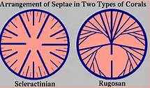
Rugose corals (Order Rugosa) are important fossil corals that were present in the Middle Ordovician to the Upper Permian. These corals had a skeleton made of the mineral calcite. The Order Rugosa contained both solitary and colonial corals with radial septa that were added in a series of four at a time (www2) (see fig. 1). The size of these rugose corals varied generally from 10-40mm in solitary forms to just 4-20mm in diameter per individual in colonial forms. There were some extremes in size with some corallites as minute as 1.5mm and some as large as 120mm in diameter. (Poty, 2010).
Tabulate corals (Order Tabulata) were present in the Early Ordovician to the Late Permian. Tabulate corals had a calcitic skeleton. These corals were all colonial. Septa were absent or greatly reduced such that they existed as ridges or spines. Tabulae were abundant in these corals (www2). Hongo & Kayanne (2009) detailed the branching network of the tabulate corals. Poty (2010) described the tabulate corals as colonies of small, narrow corallites.. The diameter of the individual corallites in tabulate colonies ranged from 0.6-15mm. Zaika (2010) outlined the anatomy of the tabulate genus Favositidae, which have a honeycomb-like structure.
Scleractinian corals (Order Scleractinia) are still seen in some forms today. Scleractinian corals were present from the Middle Triassic to the present. Scleractinian corals have an aragonitic skeleton. These corals can be colonial or solitary. Septa are added in a cyclic fashion with six being added at a time (see fig. 1). (www2). Stanley Jr. (2003) stated that there are approximately 1314 known species of living scleractinians still present today. (www1) described the scleractinian corals as hard corals, also known as hexacorals due to the septa and tentacles being found in sets of six.
References:
Campbell, N.A. & Reece, J.B., (2005 ). Biology, 7th ed. Pearson Education Inc. as Benjamin Cummings, St. Francisco.
Hongo, C. & Kayanne H., 2009. Holocene coral reef development under windward and leeward locations at Ishigaki Island, Ryukyu Islands, Japan. Sedimentary Geology, vol. 214, iss. 1-4, 62-73.
Poty,E., 2010. Morphological limits to diversification of the rugose and tabulate corals. Palaeoworld,vol. 19, iss. 3-4, 389-400.
Stanley Jr., G.D., 2003. The evolution of modern corals and their early history. Earth-Science Reviews, vol. 60, iss. 3-4, 195-225.
Zaika, Y., 2010. Structure of the corallite wall of the Upper Ordovician and Silurian Favositidae (Tabulata) and its possible use in stratigraphic correlation. Palaeoworld, vol. 19, iss. 3-4, 256-267.
www1 – http://coralreef.noaa.gov/aboutcorals/coral101/anatomy/
www2 – http://www.paleosoc.org/Corals.pdf
1 – http://eusmilia.geology.uiowa.edu/lab5b.htm
2 – http://coralreef.noaa.gov/aboutcorals/coral101/anatomy/
3 – http://www.ucmp.berkeley.edu/cnidaria/anthozoamm.html


